Oral Presentation ESA-SRB-NZSE-CaSR 2026 in conjunction with ENSA
Longitudinal ultrasound and MRI atlas of ovine fetal development (143986)
Background: Prenatal imaging is fundamental to interpreting pregnancy viability and monitoring fetal development. Ultrasound is the primary imaging modality used clinically due to its safety, accessibility, and real-time assessment. Magnetic resonance imaging (MRI) provides superior anatomical detail and soft tissue contrast but is impractical to employ regularly. Repeated imaging throughout gestation remains ethically cautious for humans and technically challenging for animal models, resulting in a lack of reference datasets. Sheep are an established translational model for human pregnancy and compatibile with clinical MRI, providing a practical opportunity to generate these datasets. This study aimed to develop a longitudinal fetal imaging atlas of sheep, integrating ultrasound and MRI to support prenatal imaginginterpretation and translational reproductive research.
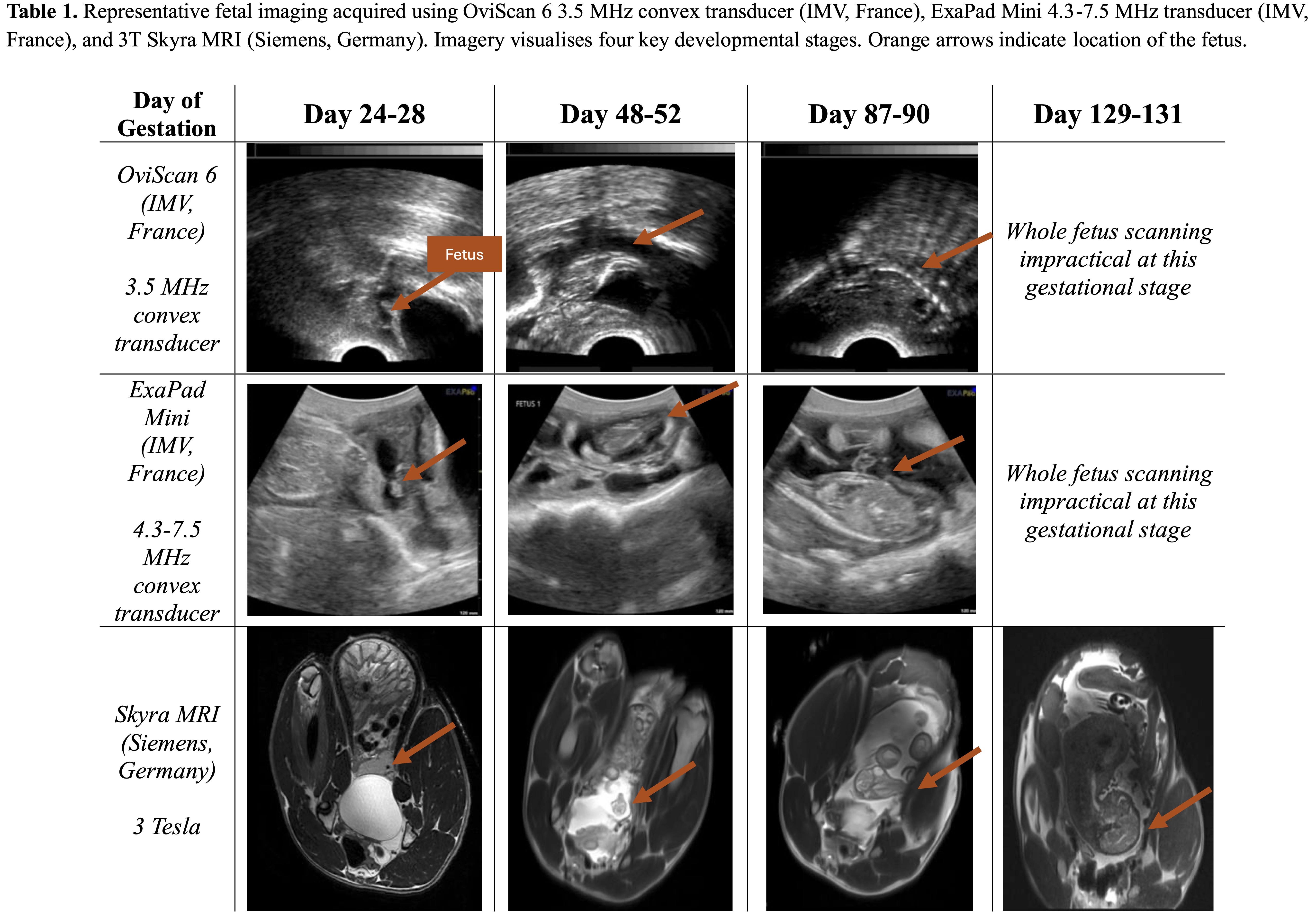
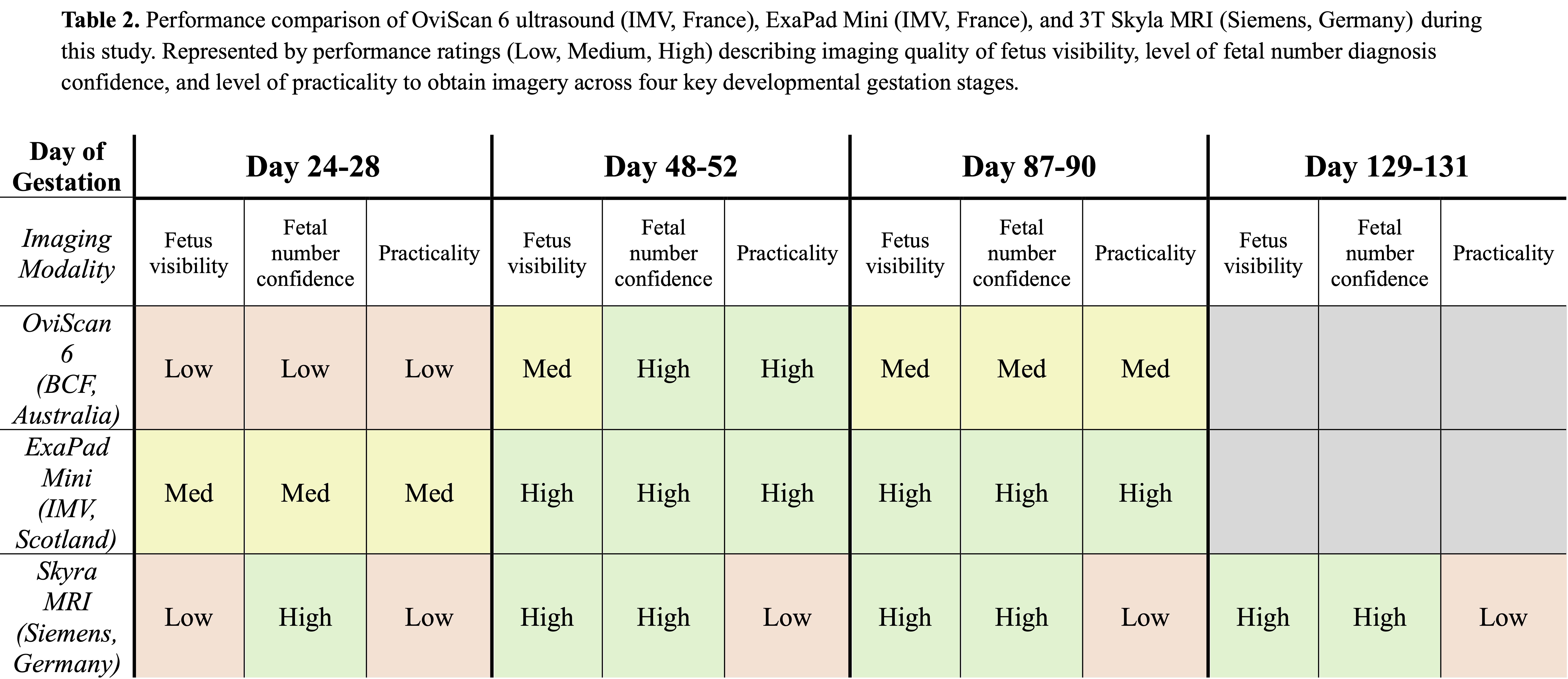
Methods: Ten synchronised, naturally mated ewes carrying singleton, twin and triplet pregnancies underwent weekly transabdominal ultrasound examinations from Days 24-90 utilising OviScan6 3.5MHz transducer (IMV, France) and ExaPad Mini 4.3-7.5MHz transducer (IMV, France). MRI examinations conducted at key developmental stages (Days 24-28, 48-52, 87-90, 129-131) using 3T Skyra MRI (Siemens, Germany). Paired ultrasound and MRI datasets were evaluated to compare contributions to prenatal imagery interpretation.
Results: Three imaging modalities provided distinct yet complementary perspectives of fetal development (Table 1,2). Lower-frequency ultrasound enabled rapid interpretation representative of sheep industry practice, while higher-frequency ultrasound provided detailed fetal anatomy and viability assessments that improved comprehension of pregnancy status. MRI offered whole-pregnancy anatomical context by confirming fetal orientation, uterine horn occupancy and spatial relationships not achievable on ultrasound. Pairing of the datasets enabled direct cross-validation between imaging modalities, with each providing context that complemented the limitations of the other.
Conclusion: This atlas establishes a normative fetal developmental reference linking ultrasound and MRI across sheep gestational stages. It provides a unique translational resource to improve prenatal image interpretation through development of educational tools and support of future reproductive biology research.