Lightning Talk + Poster ESA-SRB-NZSE-CaSR 2026 in conjunction with ENSA
ERβ Antibody Validation and Multiplex Opal Immunofluorescence Panel Development for Spatial Sex Steroid Receptor Profiling in Gonadotroph Adenomas (143881)
Background: Gonadotroph adenomas are among the most common pituitary adenomas, express estrogen receptor alpha (ERα).1 In breast cancer, estrogen receptor beta (ERβ),2 progesterone receptor (PR) and androgen receptor (AR) modulate ERα activity,3, 4 highlighting the importance of receptor interactions for hormone signalling. Such interrelations among sex steroid receptors remain unknown in the pituitary, and an adequate assay for their assessment with spatial contextualisation is critical for advancing the field. We aimed to establish a multiplex immunofluorescence panel to simultaneously quantify ERα, ERβ, PR, and AR in pituitary tumour tissue.
Methods: Chromogenic immunohistochemistry validated each antibody prior to multiplex assembly. Two ERβ antibodies, MC10 (ThermoFisher)5 and CWK-F12 (Developmental Studies Hybridoma Bank),5 were evaluated against normal breast duct epithelia. ERβ antigen retrieval was optimised by comparing pH6 and pH9 conditions. PR clones SP2 (Abcam) and 1E2 (Ventana) were validated against breast controls and quantitatively compared. AR clone 441 (Abcam) was validated on prostate control. A five-plex Opal immunofluorescence panel (PR Opal 520, AR Opal 570, ERβ Opal 690, ERα Opal 780, DAPI) was assembled. Cell segmentation and per-cell intensity quantification were performed in HALO, with quality control metrics computed in R.
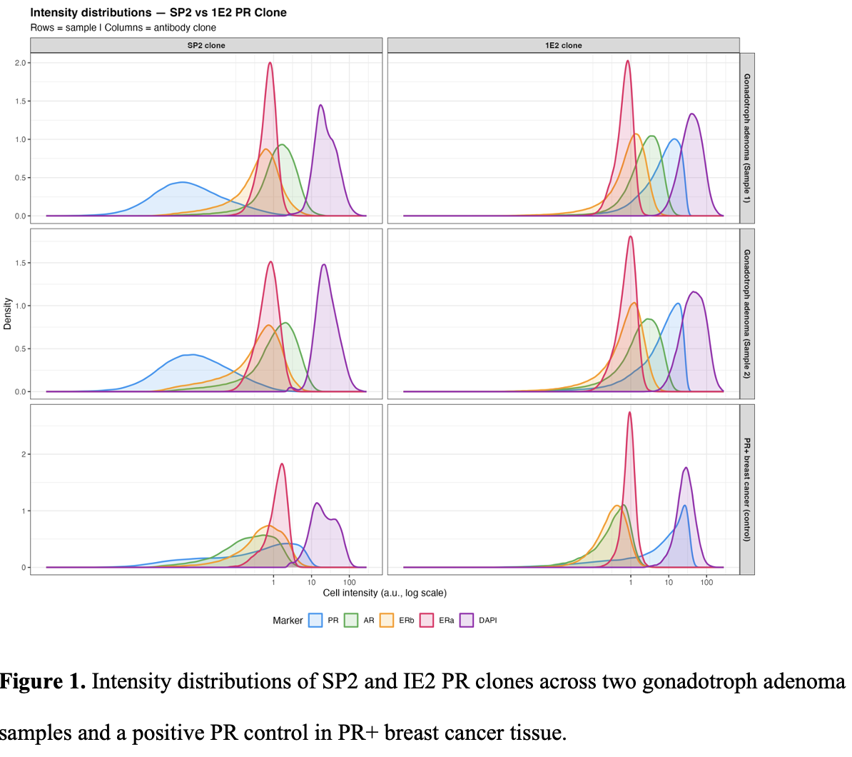
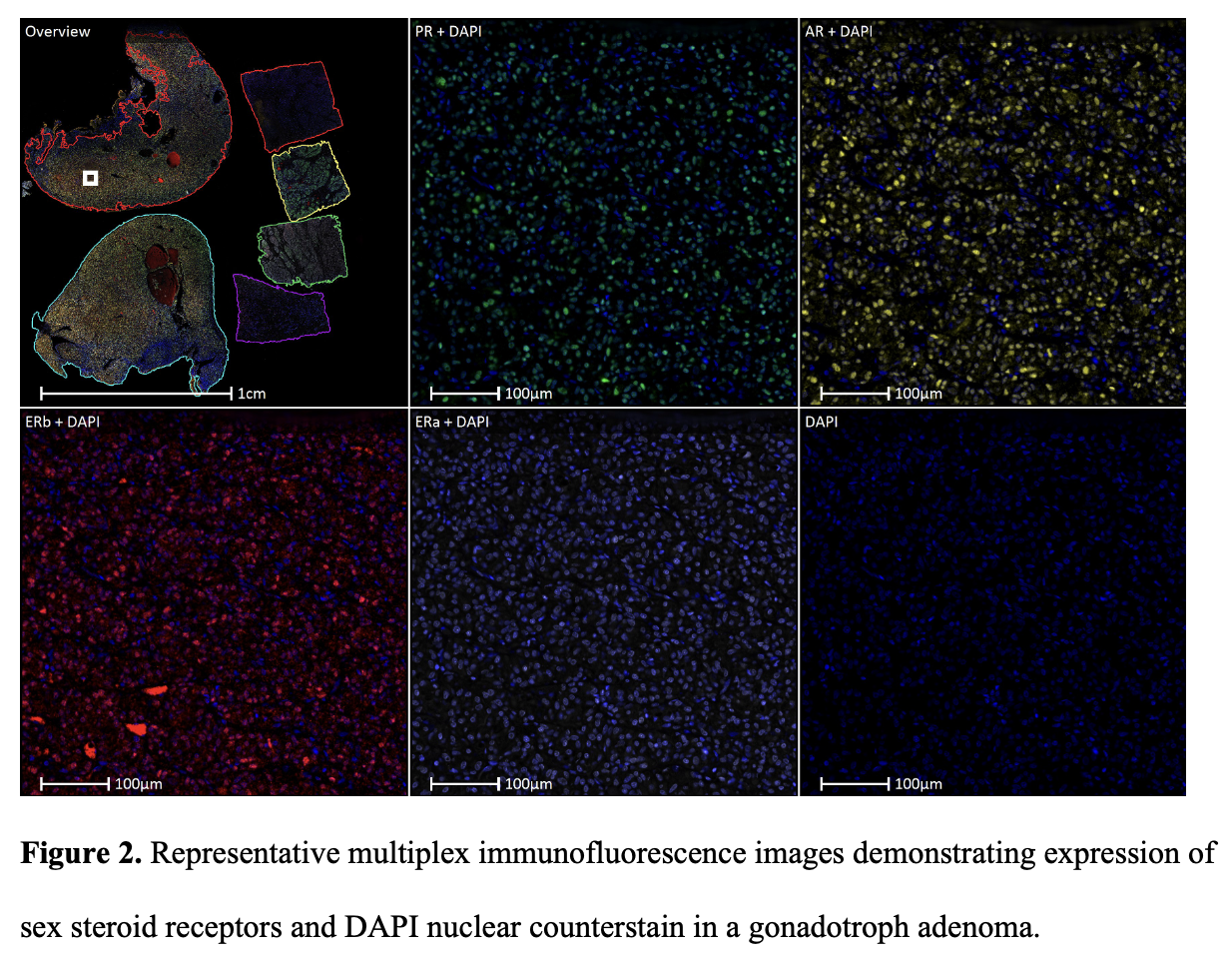
Results: CWK-F12 produced specific ERβ staining, whilst MC10 was excluded due to non-specific signal. pH6 antigen retrieval improved nuclear-specific ERβ signal versus pH9 in external control tissues. ERβ-ERα correlations were low across control tissues (r=0.09–0.37), confirming spectral channel independence. The 1E2 PR clone produced disproportionately greater improvement in pituitary than breast control tissue compared with SP2 (Figure 1), suggesting tissue-dependent PR isoform composition. All four receptors were detectable in gonadotroph adenoma tissue (Figure 2).
Conclusion: We established a validated Opal immunofluorescence panel of ERα, ERβ, PR and AR, overcoming ERβ antibody specificity, antigen retrieval and multiplex nuclear marker quantification challenges. All four receptors, including ERβ, were confirmed in gonadotroph adenomas.